Spines of tree- and ground-dwelling chameleons

Spines of tree- and ground-dwelling chameleons

Tiermedizin Science

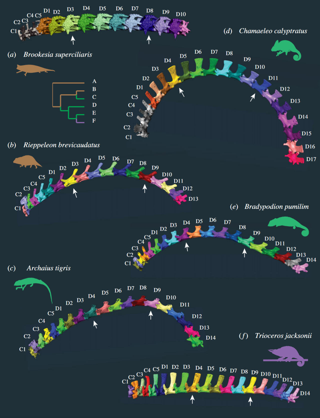
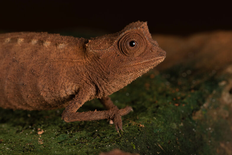
Various anatomical adaptations of the spine between ground and tree dwellers are known from mammals, especially primates. In some cases, the different vertebrae are even associated with certain movement patterns and bodily functions. In a comparative study, two scientists from New York (USA) have now investigated how the spine of ground- and tree-dwelling chameleons differs.

They measured the already existing CT scans on Morphosource.org of a total of 28 chameleons of different species. Brookesia perarmata, Brookesia superciliaris, Brookesia thieli, Palleon nasus, Rhampholeon platyceps, Rhampholeon spectrum, Rieppeleon brevicaudatus and Rieppeleon kerstenii were classified as ground dwellers. Archaius tigris, Bradypodion melanocephalum, Bradypodion pumilum, Bradypodion thamnobates, Calumma amber, Calumma brevicorne, Calumma parsonii, Chamaeleo calyptratus, Chamaeleo gracilis, hamaeleo zeylanicus, Furcifer lateralis, Furcifer pardalis, Furcifer verrucosus, Kinyongia carpenteri, Kinyongia tavetana, Kinyongia xenorhina, Nadzikambia mlanjensis, Trioceros feae, Trioceros jacksonii and Trioceros quadricornis were considered arboreal. The vertebrae were counted and the width of the lamina, length, width, height of the vertebral body, and the height of the spinous process and transverse processes on each vertebra were measured. In addition, the so-called prezygapophysial angle was determined. This is the angle of the intervertebral joint, i.e. the contact surfaces between the individual vertebrae. The measurements of ground and tree dwellers were compared and statistically evaluated. Only the vertebral column of the trunk was considered, the caudal vertebral column was left out.

First of all, the results showed that ground-dwelling chameleons generally have fewer trunk vertebrae (15 to 19) than tree-dwelling chameleons (18 to 23). The trunk spine of almost all species could be divided into the already known three areas: Cervical spine and anterior and posterior dorsal spine. A thoracic and lumbar spine as in mammals is generally not distinguished in chameleons because of the continuous ribs. Five chameleon species had four regions instead of three: they had an anterior and a posterior cervical spine, the anterior one consisting of only two vertebrae with rib processes. Six chameleon species had two additional lumbar vertebrae and one species had three transitional vertebrae in the region between the cervical and dorsal spine. In Kinyongia carpenteri, a total of five regions could be distinguished in the trunk spine: The chameleon had anterior and posterior cervical vertebrae as well as anterior and posterior dorsal vertebrae and two additional lumbar vertebrae. Brookesia perarmata was also a special case: the trunk spine of this chameleon consisted of only two regions and at the same time the smallest number of vertebrae of all species studied.

The greatest differences between ground and tree-dwelling chameleons were found in the prezygapophyseal angle (PZA) and the height of the spinous process. The intervertebral joint surfaces in the anterior dorsal vertebrae of tree-dwelling chameleons were clearly more dorsoventrally oriented and smaller than in ground-dwelling species. Several tree-dwellers showed a PZA of less than 90°. In tree-dwelling chameleons, the largest spinous processes were located at the transition from the cervical to the dorsal spine. Among the ground-dwelling species, the spinous processes were similar only in Palleon nasus. In ground-dwelling chameleons, the appearance of the spinous process varied greatly. Rieppeleon, for example, showed narrow, backward-sloping spinous processes, while the spinous processes in Brookesia were more like a kind of bone bridge than a process. Archaius tigris was an exception: The spinous processes in this chameleon hardly differed along the entire spine.

The authors conclude from the results that the anatomy of the different vertebrae is strongly related to the chameleons’ way of life and different locomotion. The intervertebral joint surfaces in tree-dwelling chameleons are probably important for climbing by supporting the function of the shoulder girdle. Reduced mobility in the mediolateral plane provides greater trunk stiffness, which facilitates climbing in arboreal dwellers. Stiffening of the axial skeleton (skull, trunk spine and thorax) is also known from tree-dwelling mammals. The larger spinous processes in larger chameleons could facilitate shoulder girdle rotation and muscle movement, resulting in increased stride length, better head support, and thus possibly easier feeding.

Morphological and functional regionalization of trunk vertebrae as an adaption for arboreal locomotion in chameleons
Julia Molnar, Akinobu Watanabe
Royal Society Open Science 10, 2023: 221509
DOI: 10.1098/rsos.221509

Illustration: Spines of different chameleon species

Long-term study on sperm collection in chameleons

Long-term study on sperm collection in chameleons

Tiermedizin Science

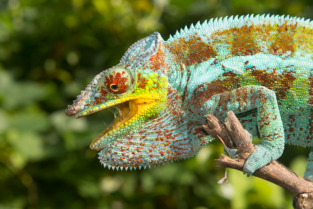
Assisted reproduction has become increasingly common in the conservation of extremely rare animals such as the Spix’s macaw or northern white rhinoceros in recent years. In reptiles, on the other hand, there have only been a few studies on assisted reproduction, and only a few on chameleons in particular. Scientists from the USA have now conducted a study on male Veiled and Panther Chameleons (Chamaeleo calyptratus and Furcifer pardalis).

At Louisiana State University, 16 males of each species were kept under standardised conditions for over a year. The panther chameleons were purchased from a US breeder, the Yemen chameleons from a dealer who had taken them from the introduced wild chameleon population in Florida. All males were kept individually in ZooMed screen cages, equipped with automatic sprinklers and artificial plants. Temperatures were around 28-29°C during the day with spots to seek higher values. 12 h UV-B irradiation per day was offered. They were fed with crickets and zophobas.

Before the start of the study, all 32 chameleons were clinically examined and parasites were treated. Only after a month of acclimatisation did the actual study begin. During the study year, all chameleons were put under anaesthesia twice a month. Each time, blood was taken from the ventral tail vein or the jugular vein to determine the testosterone concentration. Ultrasound was used to measure the size of the testicles. In addition, each time an attempt was made to obtain sperm by electroejaculation. Electroejaculation involved inserting a small metal probe into the cleaned cloaca. Each chameleon was then treated up to three times in succession with up to 15 electric shocks of 0.1/0.2/0.3 mAs. The semen collection experiments were stopped as soon as the animal ejaculated. The sperm collected was preserved and examined for ejaculate volume, presence of sperm, sperm motility, concentration, and morphology.

The results suggest that Veiled Chameleons follow a so-called prenuptial reproductive strategy under constant husbandry conditions. The testosterone concentration in the blood already increased before the sperm volume of the males had reached its maximum. The months of May, April, and June brought the best sperm volumes, the most sperm was produced by electroejaculations in the third attempt. Testicle sizes also varied throughout the year, with the largest measurements from August to December.

Panther chameleons, on the other hand, seem to follow a postnuptial reproductive strategy. In them, most sperm could only be obtained well after the highest point of testosterone concentration. The electroejaculations worked best in March, April, May and June. Much more often than in Yemen chameleons, electroejaculation in panther chameleons worked already in the first attempt. The size of the testicles also varied throughout the year, but most were largest in the months mentioned above. Together with the factors mentioned above, the volume of ejaculate, sperm concentration, sperm motility and sperm morphology also changed during the year.

The authors recommend that electroejaculation in chameleons should generally only be performed under anaesthesia. The success rate for spermatozoa in the two highest cases was 82 and 88%, which is similar to the success in other reptiles during their reproductive season. The mortality rate among the 32 animals was only 0.12% over the whole year. One panther chameleon died after 10 months during the 20th anaesthesia, after death kidney damage was detected. From the low mortality rate, the authors conclude that electroejaculation rather does not play a role in the development of kidney disease, as was suspected in other studies. However, an examination of the blood for kidney values was not carried out on any of the surviving chameleons after the study. It also remains unclear what role the lack of imitation of rainy and dry seasons during the year plays for both species and their reproductive cycle.

Characterizing the annual reproductive cycles of captive male veiled chameleons (Chamaeleo calyptratus) and panther chameleons (Furcifer pardalis)
Sean M. Perry, Sarah R. Camlic, Ian Konsker, Michael Lierz, Mark A. Mitchell
Journal of Herpetological Medicine and Surgery 33 (1), 2023, pp. 45-60
DOI: 10.5818/JHMS-D-22-00037

Unusual parasite discovered in Furcifer campani

Unusual parasite discovered in Furcifer campani

Tiermedizin Science

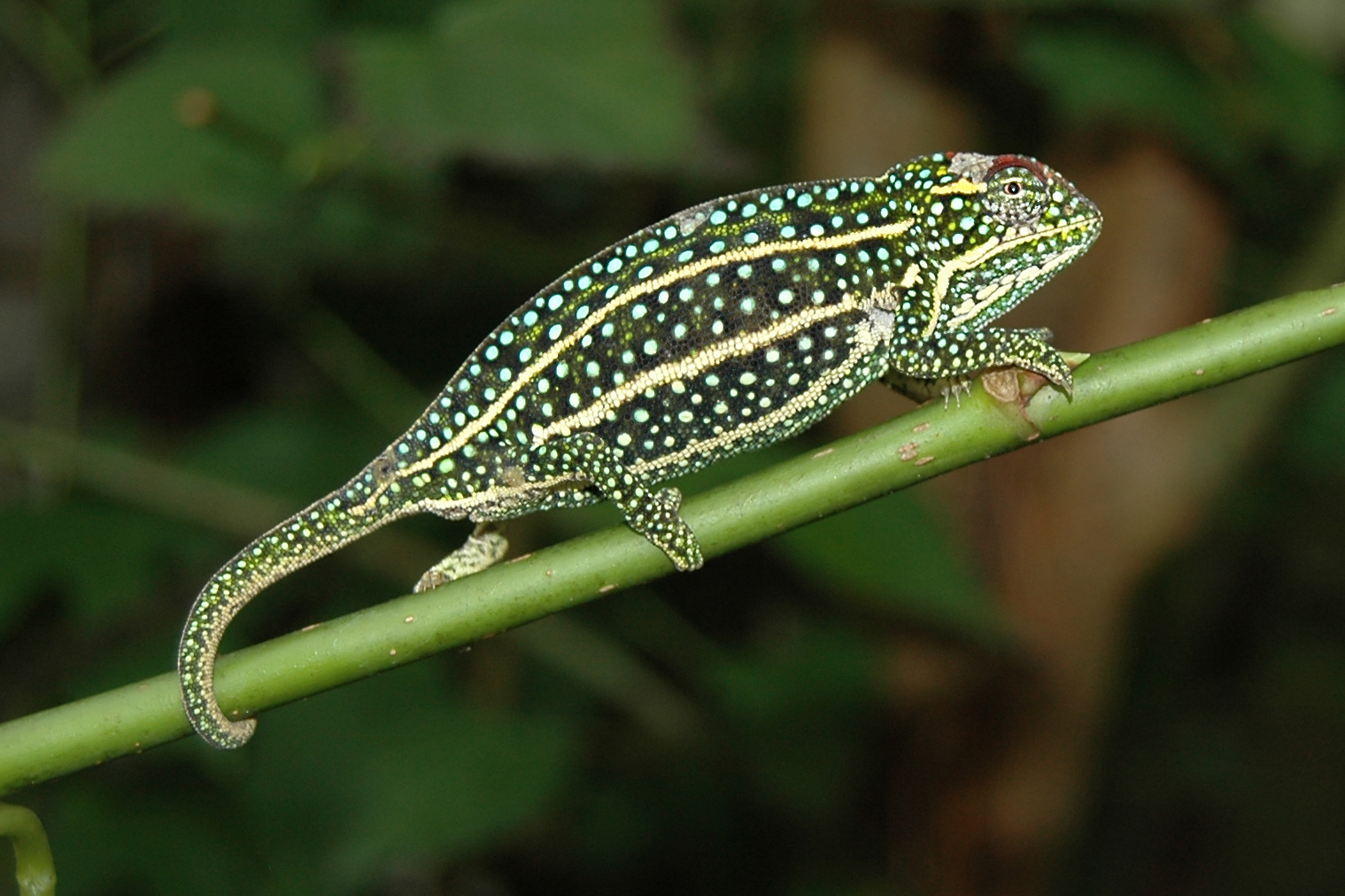
Physiologists, microbiologists and veterinarians from the USA recently described an unusual case of a parasite infestation in Furcifer campani. This is probably the new discovery of an as-yet-undescribed chameleon parasite.

In 2021, 11 Furcifer campani had been imported from Madagascar as wild-caught specimens and were kept privately. Two months after importation, unusual behaviour was noticed in one male and one female of the group. The two animals basked in the sun for unusually long periods of time, specifically seeking out temperatures of 29-30°C as well as places with enormously high UV indices compared to the other chameleons. Within the next three months, both Furcifer campani visibly lost weight, although the food supply was increased and a good food intake could be observed. At the same time, a lighter skin colouration was noticed. Faecal examinations by flotation were negative. Finally, both chameleons became lethargic, and closed their eyes during the day. A bloated abdomen and increased watery faeces were observed. Both Furcifer campani died.

Histological examination confirmed muscle atrophy and cachexia. Massive infiltration of the liver and gastrointestinal tract with large amounts of spores could be detected in both chameleons. The spores proved positive in Grocott’s methenamine-based silver stain and the PAS stain. Morphologically, the spores were classified as Dermocystidium-like. Investigations by PCR revealed a high similarity with Dermocystidium salmonis, but the exact pathogen could not be determined with certainty.

The genus Dermocystidum is a parasitic microorganism that is classified as a protist (it is neither a fungus nor an animal or plant). It is interesting that they have so far been known mainly from fish and amphibians, occasionally also from mammals. So far, not a single case of infestation with Dermocystidium has been described from reptiles. It could therefore be an undescribed, new species that is possibly even chameleon-specific. Effective therapy is not yet known.

A unique disease presentation associated with a mesomycetozoean-like organism in the jeweled chameleon (Furcifer campani)
Michael Nash, Emily A. McDermott, Ashley K. McGrew, Juan Muñoz, Dayna Willems
Journal of Herpetological Medicine and Surgery, February 2023
DOI: 10.5818/JHMS-D-22-00033

Minimally invasive methods for obtaining DNA samples from chameleons

Minimally invasive methods for obtaining DNA samples from chameleons

Tiermedizin Science

To reliably identify or compare chameleon species, genetic samples of the animals concerned are necessary. Traditionally, scientists have used organ or muscle samples from euthanized chameleons in museum collections or – less commonly – cut tail tips or blood samples from living chameleons. Researchers at the American College in Athens, Greece, have studied whether more minimally invasive methods would also be a good alternative.

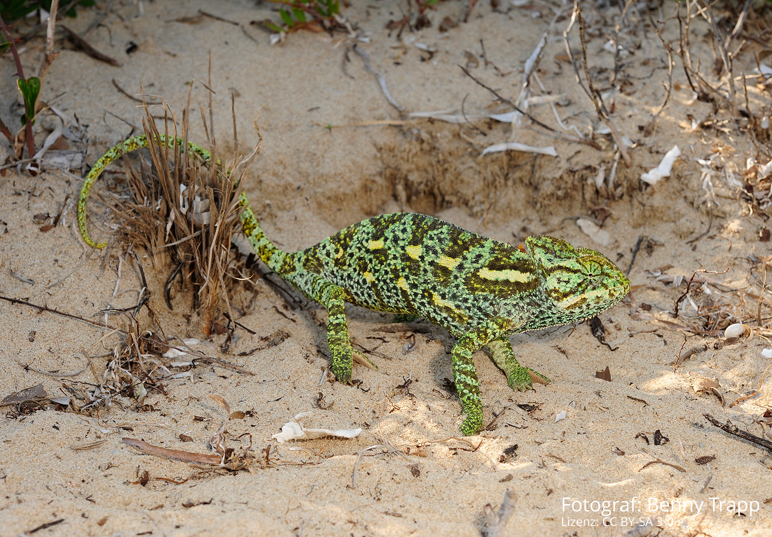
They sampled 23 Chamaeleo africanus in the area of the lagoon of Pylos (Divari wetland between Gialova and the bay of Voidokilia) in the Peloponnese in Greece using buccal swabs. This involves running a sterile swab on the inside of the cheek through the chameleon’s mouth for six seconds. Blood was taken from the ventral tail vein of eight other Chamaeleo africanus for comparison. Sampling took less than a minute. Afterward, the chameleons were returned to where they were found. The swabs were transported refrigerated in a special buffer solution in Eppendorf cups and then frozen.

In the laboratory, the researchers were able to extract both nuclear and mitochondrial DNA from all the swabs. However, the quantity and quality of the DNA extracted were lower than in the blood samples. For most applications such as PCR amplification and gene sequencing, however, the scientists said the quantity was sufficient. In terms of invasiveness and destructiveness, buccal swabbing is certainly preferable to killing or injuring individual chameleons. Studies on other reptiles suggest that rapid freezing is not mandatory either – in the field, a functioning cool chain could become a problem in many chameleons’ countries of origin. The current study advises against ethanol as a fixing solution; the buffer solutions used lead to better results.

Buccal swabbing appears to be less applicable for cases where additional material for future studies might be preserved, for example when describing new species, or when sequencing the entire genome. However, the method is certainly a good alternative, especially for particularly small chameleon populations where lethal sampling could already significantly limit the breeding pool.

Buccal swabs as an effective alternative to traditional tissue sampling methods for DNA analyses in Chamaeleonidae
Maria Koutsokali, Christina Dianni and Michael Valahas
Wildlife Biology
DOI: 10.1002/wlb3.01052

Parasite treatment in leaf chameleons

Parasite treatment in leaf chameleons

Tiermedizin

Last weekend, the autumn meeting of the AG Amphibien- und Reptilienkrankheiten (working group on amphibian and reptile diseases) took place. With over 500 members, the AG ARK is one of the strongest sub-groups of the DGHT and at the same time the largest association of veterinarians for amphibians and reptiles in Europe. Accordingly, the autumn meeting in Münster was fully booked as usual. Besides the main topic of the meeting, Asian turtles, one veterinarian presented a case report on parasite treatment in leaf chameleons.

In this case, 7.5 subadult and adult Brookesia stumpffi (5 wild-caught, 7 German offspring from different husbandries) were acquired for a breeding project. Fecal examinations of all terrestrial chameleons were carried out. Masses of Choleoeimeria spp., presumably Choleoeimeria brookesiae, were found in flotation and native preparations of almost all terrestrial chameleons. In addition, there were isolated co-infections with Heterakis spp. and trematodes. Strongylid-like eggs with thin shells and larvae as well as adult nematodes were found in the feces of several animals. A treatment protocol with Baycox 50 mg/ml (Elanco Animal Health, Rathausplatz 12, 61352 Bad Homburg, Germany, active ingredient toltrazuril) on days 1, 7, and 14 and Panacur 10% (Intervet Germany, Feldstraße 1a, 85716 Unterschleissheim, Germany, active ingredient fenbendazole) on day 3 and 13 proved successful. The diluted solutions were given into the mouth with a 100 µl pipette. Fecal examinations at the beginning of quarantine and on days 14, 28, and 42 after treatment were suggested as a practical protocol for veterinarians.

The biggest problem during treatment was reinfection with coccidial oocysts from the environment. The leaf chameleons reinfected themselves, among other things, via left feeders and fecal remains on climbed gauze and living plants. Successful quarantine was finally achieved under the following parameters: individual keeping without visual contact in separate terrariums, daily exchange of kitchen paper on the floor and a freshly cut elder branch, use of new gloves for each chameleon, slow-moving food from bowls disinfected daily in boiling water, weekly disinfection with ready-to-use Interkokask® (Albert Kerbl GmbH, Felizenzell 9, 84428 Buchbach, active ingredient chlorocresol). Extremely strict compliance with all cleaning and disinfection measures was necessary.

Parasite treatment in leaf chameleons (Brookesia stumpffi)
Dr. Alexandra Laube
Proceedings of the 57th Workshop of the WG Amphibian and Reptile Diseases, Focus: Asian Turtles
Münster, 04 – 06 November 2022