Scientists at the University of Veterinary Medicine Montréal (France) recently analysed the causes of death in chameleons kept in zoos between 2011 and 2022. The Zoological Information Management System (ZIMS) was used to search for zoos that currently keep chameleons or have kept them since 2011. Questionnaires were sent to a total of 245 zoos. The questionnaires asked about the number, species and sex of chameleons kept, as well as selected husbandry conditions (coolest and warmest temperatures, humidity, feeding) and dissection results.
Around 1000 chameleons of 36 different species are currently kept in zoos worldwide. 65 of the zoos surveyed took part in the study, 48 of which regularly carried out dissections on chameleons. However, only 29 of the participating zoos were able to provide dissection results. A total of 412 pathological findings from 14 different chameleon species were analysed. Among the species kept were Brookesia stumpffi, Brookesia superciliaris, chameleons of the genus Brookesia without species identification, Calumma parsonii, Chamaeleo calyptratus, Chamaeleo chamaeleon, Furcifer lateralis, Furcifer oustaleti, Furcifer pardalis, Rieppeleon brevicaudatus, Trioceros melleri, Trioceros montium and Trioceros quadricornis. Panther chameleons were kept most frequently (226 specimens).
The statistical analysis showed that most of the chameleons in the participating zoos died of infectious diseases (46.8%). Infectious diseases included septicaemia, but also inflammation of the oral cavity, lungs, liver, kidneys and intestines. Almost 20% of the infectious diseases were in the area of the oral cavity. The most common bacteria were Enterococcus and Pseudomonas. Among the fungi, Nannizziopsis including CANV, Fusarium and Metarhizium were represented. A good third of the necropsy reports also indicated parasitoses, with these occurring both as a cause of death and as an incidental finding. Coccidia and trematodes as well as various nematodes were often present. The second most common cause of death in the participating zoos was non-infectious kidney diseases (11.4%). This was closely followed by diseases of the reproductive tract, including egg loss and egg yolk coelomitis, which accounted for 10.7% of cases.
Contrary to the authors’ initial assumption, there was no correlation between the surveyed husbandry parameters in the cages and the incidence of kidney disease. Basically, there was a tendency towards an increased incidence of kidney disease in countries where the average humidity was generally lower.
Evaluation of mortality causes and prevalence of renal lesions in zoo-housed chameleons: 2011-2022
Amélie Aduriz, Isabelle Lanthier, Stéphane Lair, Claire Vergnau-Grosset
Journal of Zoo and Wildlife Medicine 55(2), 2024
DOI: 10.1638/2023-0023
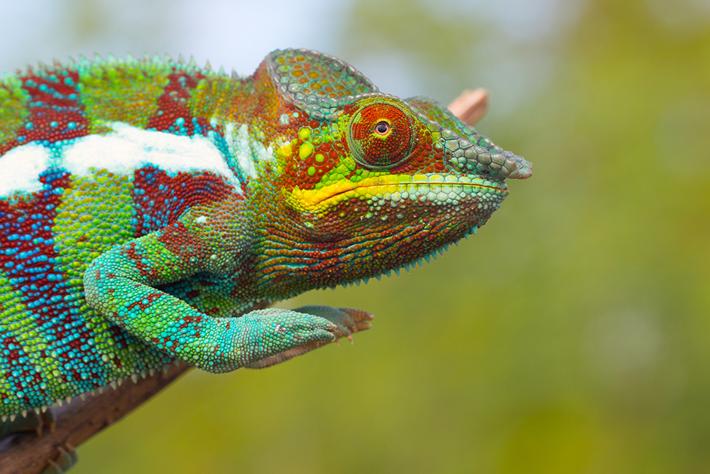
Photo: Panther chameleon in Madagascar, photographed by Alex Negro


