Sex chromosomes in chameleons

Sex chromosomes in chameleons

Science

Which sex chromosomes are present in chameleons has so far been studied rather sparsely. The Madagascan chameleon genus Furcifer is known to have Z and W chromosomes, although sometimes several Z chromosomes occur, so-called neo-sex chromosomes. Recently in the Czech Republic, scientists examined this deeper.

Blood and tissue samples were taken from 13 chameleons to isolate DNA. The animals sampled included one male and one female each of the species Brookesia therezieni, Calumma glawi, Calumma parsonii, Chamaeleo calyptratus, Furcifer campani, Furcifer labordi, Furcifer lateralis, Furcifer oustaleti, Furcifer pardalis, Furcifer rhinoceratus, Furcifer viridis, Kinyongia boehmei and Trioceros johnstoni. Only in Furcifer oustaleti were two females sampled. Subsequently, the Z1 chromosomes of the panther chameleons and the Z and W chromosomes were analysed by microdissection. Gene coverage analyses were performed for carpet and panther chameleons. In addition, qPCRs were performed to compare the homology of the Z chromosomes.

The results show that the morphology of the Z1 chromosomes of panther chameleons corresponds to the Z chromosome of the entire genus Furcifer. The Z1 chromosome of panther chameleons thus corresponds to the Z chromosome of Furcifer oustaleti. The Z2 chromosome of panther chameleons, on the other hand, is a neo-sex chromosome. Both the Z and W chromosomes in Furcifer oustaleti are probably pseudautosomal. 42 genes have been described as specific for the W chromosome.

A total of 16,947 genes were identified in Furcifer lateralis and 16,909 genes in Furcifer pardalis. The ratio of the number of genes between females and males is 0.35 and 0.65 for the two species. In panther and carpet chameleons, most of the genes on the W and Z chromosomes were found to be the same, with relatively few genes found only on the W chromosome. This finding is surprising, as the researchers had actually expected that the heterochromatic W in Furcifer species would have lost most of its genes compared to the Z chromosome.

The sex chromosomes of the genus Furcifer probably evolved at least 20 million years ago, which roughly corresponds to the time when the species Furcifer campani split off from the other Furcifer species.

Heteromorphic ZZ/ZW sex chromosomes sharing gene content with mammalian XX/XY are conserved in Madagascan chameleons of the genus Furcifer
Michail Rovatsos, Sofia Mazzoleni, Barbora Augstenová, Marie Altmanová, Petr Velenský, Frank Glaw, Antonio Sanchez, Lukáš Kratochvíl
Scientific Reports 14, 2024: 4898.
DOI: 10.1038/s41598-024-55431-9

New case reports on hemipenes amputation

New case reports on hemipenes amputation

Tiermedizin

The University of Sofia (Bulgaria) has published a new paper with several case reports involving chameleons. The authors describe 16 cases of different lizards that suffered a hemipenis prolapse and their treatment.

The lizards included a panther chameleon (Furcifer pardalis) and two Veiled Chameleons (Chamaeleo calyptratus). All three patients were presented to the veterinarians with bilateral hemipenes prolapse. Initially, the prolapses were bathed in 20% dextrose solution, after which the hemipenes were manually repositioned. However, the prolapses then recurred, so surgery was the final solution. Under general and local anaesthesia administered intramuscularly, the hemipenes were removed, the wound sutured and the remaining small stump repositioned in the respective hemipenes pocket. Meloxicam was administered as an analgesic once a day for 5 days after the operation. Only lizards in which the surgical field appeared to be dying off during the follow-up examinations were given antibiotics for 10 days.

Hemipenectomy in leopard geckos, chameleons and bearded dragons
Seven Mustafa & Iliana Ruzhanova-Gospodinova
Tradition and Modernity in Veterinary Medicine, 2024
DOI: nicht vorhanden

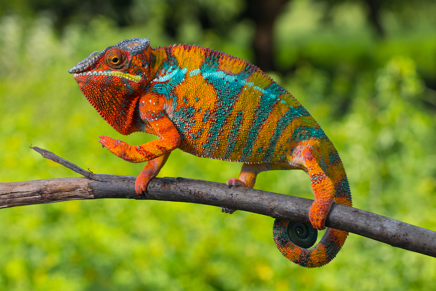
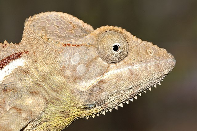
Photo: Panther chameleon, photographed by Alex Laube in Madagascar

Lecture for vets on prophylaxis for chameleons

Lecture for vets on prophylaxis for chameleons

Tiermedizin Webinars

On 18 November 2023, the spokesperson of the AG Chameleons will give a lecture for veterinary colleagues on which prophylactic measures are possible and useful in veterinary practice. To a large extent, prophylaxis includes keeping the chameleons in the terrarium itself, so a large part of the lecture will deal with what needs to be considered when keeping chameleons and which common mistakes still lead to husbandry-related diseases or injuries in chameleons. She will also discuss the collection of the so-called minimum database during the annual check-up in the veterinary practice and show examples of diseases recognised early and late. A Q&A session directly after the respective presentations rounds off the short excursion into prophylaxis in chameleons.

Registration for the conference is also possible for non-DGHT members who are veterinarians – see the link below to the mVet conference platform.

Dr. Alexandra Laube Prophylaxis for chameleons – is it possible and if so, how?
59. Conference of DGHT working group amphibian and reptile diseases (AG ARK)
Online

Photo: Calumma amber in the Montagne d’Ambre, Madagascar, photographed by A. Laube

Aggressive fungal pathogen discovered in panther chameleons

Aggressive fungal pathogen discovered in panther chameleons

Tiermedizin Science

Fungi of the genera Nannizziopsis and Paranannizziopsis have long been known to cause severe skin diseases in various reptiles. These include species feared in herpetocolture such as CANV (Chrysosporium Anamorph of Nannizziopsis vriesii) and Nannizziopsis dermatitidis, which are apparently obligate pathogens. Now, a similar skin fungus has been detected in panther chameleons in Florida, USA.

Nine adult panther chameleons (Furcifer pardalis) were taken from a wild population in Florida. They were first housed in groups of two or three chameleons in screen cages with natural and artificial plants at a private keeper. A ReptiSun 5.0 lamp and a conventional light bulb were used. The animals were fed with crickets and zophobas every second day and supplemented with vitamins and calcium. All nine panther chameleons, plus a tenth that was captured later, were finally given to the United States Department of Agriculture (USDA) for a series of experiments. They were kept individually in steel aviaries outdoors.

Subsequently, the eight remaining panther chameleons were also examined. In fact, all but one of the chameleons were found to have either missing claws or swelling of the hands and feet, small skin wounds, circumferential proliferation on the body and/or yellow and black skin lesions. Fungal PCR was no longer carried out, but infection with the same pathogen was suspected. All nine panther chameleons still alive were treated with 25 mg/kg terbinafine and 5 mg/kg voriconazole, both given orally once daily.

After six weeks, the panther chameleons were examined again. The skin lesions were still present, in two animals the hand and foot swellings had decreased. After eleven to twelve weeks of treatment, all symptoms had disappeared in seven chameleons. The skin lesions had developed into scars. Only two chameleons still showed swelling in the foot area, but less than at the beginning of treatment. After 14 weeks of therapy, another panther chameleon died. The autopsy revealed kidney and organ damage as the cause of death. Since the contribution of the medication used to the death of the chameleon could not be ruled out, the therapy was terminated in week 15 for six of the eight panther chameleons. The two panther chameleons that still showed swelling of the feet were treated for another two weeks.

This case report is the first detection of Paranannizziopsis australasiensis in chameleons. So far, this fungal pathogen has only been found in green iguanas and Eastern bearded dragons in herpetoculture and in Tuataras, skinks, geckos, and snakes in nature. It remains unclear where the panther chameleons became infected. Three of the screen cages used in the initial private husbandry had previously been inhabited by Veiled Chameleons (Chamaeleo calyptratus) and Knight Anoles. The private keeper had disinfected the terrariums with chlorine bleach. The rest of his reptile population showed no skin lesions. The aviaries used later at the USDA had been empty for years and had previously only been inhabited by small birds. It is possible that the fungal pathogen had been introduced via potted plants from nurseries that regularly observe native reptiles on the premises. However, Paranannizziopsis australasiensis has not been found in any other wild reptile species in Florida to date.

The most likely scenario seems to be that the chameleons were already infected before they were caught in Florida, but the disease only broke out later. The original animals of the population could have been infected by bearded dragons in the pet trade a good decade ago. A latent infection with a late onset is supported by the fact that most of the skin lesions in this case report were found in winter, after temperatures had dropped below 10°C. Moreover, before the onset of symptoms, so-called “thermal limit trials” were carried out, in which the animals were briefly exposed to extreme temperatures of up to 45°C and 6°C. Another chameleon from the same population was caught at a later time and also developed skin lesions, which indeed suggests an infected population in Florida.

A free-ranging chameleon population infected with Paranannizziopsis australasiensis could pose a huge risk to native reptiles. The fungal pathogen is known to be highly infectious and aggressive. In addition, free-ranging panther chameleons in Florida are now being captured by dealers and sold to private owners, which could result in the spread of the disease in private reptile populations. Further research is urgently needed to clarify the extent of the current occurrence of Paranannizziopsis australasiensis in Florida, both in herpetoculture and in the wild.

Dermatomycosis caused by Paranannizziopsis australasiensis in nonnative panther chameleons (Furcifer pardalis) captured in Central Florida, USA
Natalie M. Claunch, Colin M. Goodman, Madison Harman, Mariaguadalupe Vilchez, Savanna D. Smit, Bryan M. Kluever, James F.X. Wellehan, Robert J. Ossiboff, Christina M. Romagosa
Journal of Wildlife Diseases (4), 2023
DOI: 10.7589/JWD-D-22-00018

Unusual parasite discovered in Furcifer campani

Unusual parasite discovered in Furcifer campani

Tiermedizin Science

Physiologists, microbiologists and veterinarians from the USA recently described an unusual case of a parasite infestation in Furcifer campani. This is probably the new discovery of an as-yet-undescribed chameleon parasite.

In 2021, 11 Furcifer campani had been imported from Madagascar as wild-caught specimens and were kept privately. Two months after importation, unusual behaviour was noticed in one male and one female of the group. The two animals basked in the sun for unusually long periods of time, specifically seeking out temperatures of 29-30°C as well as places with enormously high UV indices compared to the other chameleons. Within the next three months, both Furcifer campani visibly lost weight, although the food supply was increased and a good food intake could be observed. At the same time, a lighter skin colouration was noticed. Faecal examinations by flotation were negative. Finally, both chameleons became lethargic, and closed their eyes during the day. A bloated abdomen and increased watery faeces were observed. Both Furcifer campani died.

Histological examination confirmed muscle atrophy and cachexia. Massive infiltration of the liver and gastrointestinal tract with large amounts of spores could be detected in both chameleons. The spores proved positive in Grocott’s methenamine-based silver stain and the PAS stain. Morphologically, the spores were classified as Dermocystidium-like. Investigations by PCR revealed a high similarity with Dermocystidium salmonis, but the exact pathogen could not be determined with certainty.

The genus Dermocystidum is a parasitic microorganism that is classified as a protist (it is neither a fungus nor an animal or plant). It is interesting that they have so far been known mainly from fish and amphibians, occasionally also from mammals. So far, not a single case of infestation with Dermocystidium has been described from reptiles. It could therefore be an undescribed, new species that is possibly even chameleon-specific. Effective therapy is not yet known.

A unique disease presentation associated with a mesomycetozoean-like organism in the jeweled chameleon (Furcifer campani)
Michael Nash, Emily A. McDermott, Ashley K. McGrew, Juan Muñoz, Dayna Willems
Journal of Herpetological Medicine and Surgery, February 2023
DOI: 10.5818/JHMS-D-22-00033

Parasite treatment in leaf chameleons

Parasite treatment in leaf chameleons

Tiermedizin

Last weekend, the autumn meeting of the AG Amphibien- und Reptilienkrankheiten (working group on amphibian and reptile diseases) took place. With over 500 members, the AG ARK is one of the strongest sub-groups of the DGHT and at the same time the largest association of veterinarians for amphibians and reptiles in Europe. Accordingly, the autumn meeting in Münster was fully booked as usual. Besides the main topic of the meeting, Asian turtles, one veterinarian presented a case report on parasite treatment in leaf chameleons.

In this case, 7.5 subadult and adult Brookesia stumpffi (5 wild-caught, 7 German offspring from different husbandries) were acquired for a breeding project. Fecal examinations of all terrestrial chameleons were carried out. Masses of Choleoeimeria spp., presumably Choleoeimeria brookesiae, were found in flotation and native preparations of almost all terrestrial chameleons. In addition, there were isolated co-infections with Heterakis spp. and trematodes. Strongylid-like eggs with thin shells and larvae as well as adult nematodes were found in the feces of several animals. A treatment protocol with Baycox 50 mg/ml (Elanco Animal Health, Rathausplatz 12, 61352 Bad Homburg, Germany, active ingredient toltrazuril) on days 1, 7, and 14 and Panacur 10% (Intervet Germany, Feldstraße 1a, 85716 Unterschleissheim, Germany, active ingredient fenbendazole) on day 3 and 13 proved successful. The diluted solutions were given into the mouth with a 100 µl pipette. Fecal examinations at the beginning of quarantine and on days 14, 28, and 42 after treatment were suggested as a practical protocol for veterinarians.

The biggest problem during treatment was reinfection with coccidial oocysts from the environment. The leaf chameleons reinfected themselves, among other things, via left feeders and fecal remains on climbed gauze and living plants. Successful quarantine was finally achieved under the following parameters: individual keeping without visual contact in separate terrariums, daily exchange of kitchen paper on the floor and a freshly cut elder branch, use of new gloves for each chameleon, slow-moving food from bowls disinfected daily in boiling water, weekly disinfection with ready-to-use Interkokask® (Albert Kerbl GmbH, Felizenzell 9, 84428 Buchbach, active ingredient chlorocresol). Extremely strict compliance with all cleaning and disinfection measures was necessary.

Parasite treatment in leaf chameleons (Brookesia stumpffi)
Dr. Alexandra Laube
Proceedings of the 57th Workshop of the WG Amphibian and Reptile Diseases, Focus: Asian Turtles
Münster, 04 – 06 November 2022